Back pain with osteochondrosis
Osteochondrosis is a chronic disease that affects the intervertebral discs and cartilage tissue. It is widespread and mainly affects people over the age of forty, although the first symptoms can appear as early as the age of twenty or thirty. In most cases, back pain is caused by osteochondrosis.
The human spine consists of several dozen vertebrae. To prevent the sensitive vertebrae from rubbing against each other, there are so-called intervertebral discs between each pair, which perform a shock-absorbing function. With osteochondrosis, blood circulation in the spine is disrupted, which leads to metabolic disorders, and these processes affect the intervertebral discs.
When the intervertebral discs thin out during the course of the disease and cease to perform their functions, we can talk about the development of a disease called spinal osteochondrosis. Osteochondrosis of the spine can develop in a separate part - the cervical, thoracic or lumbosacral region, or affect the entire spine.

NORMAL SPINE
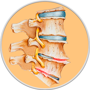
OSTEOCHONDROSIS
The diagnosis of osteochondrosis is established based on the patient's complaints, objective examination data, and X-ray examination. X-ray remains the simplest and most widely available method of examining joints to assess anatomical changes in bone structure in osteochondrosis. The method of X-ray diagnostics allows you to assess both the entire spinal column as a whole and its individual sections: cervical, thoracic, and lumbosacral. X-rays allow you to determine the localization of the process, establish its nature and severity, as well as identify the consequences of osteochondrosis, such as narrowing of the spinal canal, the presence of osteophytes, and signs of spondyloarthrosis. X-rays are usually performed in two projections: direct (in the supine position) and lateral or oblique projection. No special preparation is required for the examination, except for the lumbar spine X-ray, which requires bowel cleansing.
For a more in-depth assessment of the condition of the spinal column and its functional capabilities, it is necessary to perform an X-ray of the spine with functional tests, that is, while performing special exercises.
With the progression of osteochondrosis, pinched nerve roots, or suspected intervertebral hernia, magnetic resonance imaging is used for accurate diagnosis, which allows you to detect changes in the soft tissues surrounding the spinal column.