The effects of sports on joints

1. Football/Hockey. Post-traumatic knee osteoarthritis
2. Skiing/snowboarding. Post-traumatic ankle osteoarthritis
3. Tennis/Golf. Lateral Epicondylitis
Sports injuries are constant companions not only of professional athletes. The fashion for a healthy lifestyle and the widespread enthusiasm for various types of physical activity, in addition to the undeniable health benefits, entail increased risks of injury and joint diseases. If earlier arthrosis was characteristic only for people of advanced age, these days these degenerative-dystrophic joint diseases are increasingly found in young, active and, it would seem, people who monitor their health. Why does this happen and what should be feared when doing this or that sport?
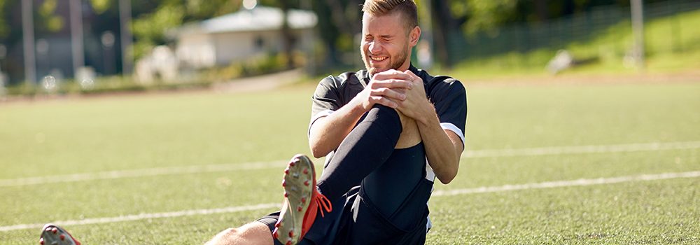
Football/Hockey
Post-traumatic osteoarthritis of the knee joint

The stands froze in tense anticipation, a dangerous moment – the attacker comes face to face with the opponent! A tackle, a kick… and a goal?! No! – A torn cruciate ligament in the knee joint. Not only professional football and hockey players, but also amateur players face this injury.
Treatment of ligament rupture involves prolonged immobilization, that is, immobilization of the knee joint for several months, which in turn threatens the development of other joint problems. The fact is that against the background of functional "inactivity" the cartilage tissue is not renewed, and therefore does not produce a sufficient amount of synovial fluid. This condition becomes a trigger for the development of post-traumatic arthrosis.
According to statistics, rupture of the cruciate ligaments of the knee joint in 20-60% cases leads to the development of arthrosis.
Post-traumatic osteoarthritis is a disease in which the cartilage lining the joint surfaces is destroyed. Damage to the cartilage causes a secondary inflammatory process in the joint, which further destroys the cartilage plate. The vicious circle is closed.
Symptoms of post-traumatic arthrosis
Like any other osteoarthritis, post-traumatic osteoarthritis of the knee joint is manifested by pain and limited mobility.
Pain in the initial stage of arthrosis is moderate and occurs during movement. A characteristic feature of the pain syndrome in arthrosis is the appearance of pain when climbing or descending stairs. Another typical sign of arthrosis is “starting pain” – the appearance of unpleasant sensations in the knee at the moment of starting movements, for example, after a night's sleep.
As osteoarthritis progresses, both rest pain and night pain appear. Constant aching pain in the knee joint, which does not subside even at rest, disrupts sleep and significantly reduces a person's quality of life.
In addition to pain, limitation of mobility in the joint is also important in the symptoms of arthrosis. As the cartilage breaks down, it is replaced by denser bone tissue, which gradually reduces the amplitude of movements in the joint, and later, due to the bone fusion of the articulating surfaces, ankylosis develops - complete immobility of the joint.
Diagnosis of arthrosis
If a professional or amateur athlete develops joint pain a few months after a knee injury, the most likely diagnosis is osteoarthritis. To confirm his suspicions, the doctor will order an X-ray, the main screening method for diagnosis. An X-ray image shows osteoarthritis:
- narrowing of the joint space;
- the presence of bone growths – osteophytes;
- compaction of bone tissue under the cartilage plate – subchondral sclerosis.
However, X-ray examination does not always answer all the questions of the diagnostician. A more informative diagnostic method is magnetic resonance imaging (MRI). Tomography allows you to thoroughly study the condition of the ligaments, menisci, articular cartilage and capsule.
Arthroscopy has been widely used in recent years for accurate diagnosis. This is the visualization of the joint cavity using a camera inserted into the joint cavity.
Treatment of post-traumatic arthrosis
The basic principles of treating joint diseases are usually well known to those who have experienced a knee injury. Unloading the knee with specially selected orthoses, physiotherapy courses, special gymnastic exercises, as well as non-steroidal anti-inflammatory drugs (NSAIDs) to eliminate pain.
In recent years, injectable chondroprotectors based on a bioactive concentrate obtained from small marine fish have become widespread. The drugs contain components that promote the regeneration of cartilage tissue and allow you to achieve long-term results, rather than temporary relief of pain.
Skiing/snowboarding
Post-traumatic ankle arthritis

Skiing or snowboarding enthusiasts are eagerly awaiting the arrival of snow in the mountains. Bright emotions, drive, and adrenaline await. Everything is fine, except for a small spoonful of tar – the danger of injury. Most often, skiers and snowboarders are threatened by ankle fractures. And this is always prolonged immobilization and secondary destruction of the cartilage lining the articular surfaces of the talus and tibia. This is how post-traumatic ankle arthritis develops.
Osteoarthritis in a skier or snowboarder is not always associated with injury; it is often caused by the increased stress on the ankle joint that accompanies winter sports.
Symptoms and diagnosis of osteoarthritis
The first symptoms of the disease usually appear a few months after the end of the season. Most often, even before the appearance of pain in the ankle joint, attention is drawn to the crunching sound when moving the joint and swelling of the ankles and feet, associated with impaired microcirculation. Gradually, pain in the ankle joins during physical exertion, limping appears, the person notices that he cannot bend or unbend the foot as actively as before.
Only a doctor can make an accurate diagnosis based on the results of an X-ray examination. A typical X-ray picture of ankle osteoarthritis reveals:
- reduction in the height of the joint gap between the talus and tibia;
- the appearance of bone growths along the edges of the articular surfaces;
- increasing bone density in the area directly under the cartilage.
Treatment
The principles of treatment of ankle arthrosis are the same as in the treatment of arthrosis of other localizations. During the exacerbation, it is necessary to provide rest to the damaged joint (stop sports, wear an orthosis or bandage the joint), eliminate pain syndrome (NSAIDs are traditionally used for this purpose) and prevent further destruction of cartilage. For this, drugs that promote the regeneration of cartilage tissue - chondroprotectors - are used in basic therapy. This is the only group of modern drugs that directly affects the cause and pathogenesis of arthrosis, and not just eliminates symptoms.
Modern chondroprotectors based on bioactive concentrate contain a complex of biologically active substances (chondroitin sulfate, amino acids, trace elements), thanks to which it is possible to:
- Accelerate the process of cartilage renewal;
- Activate the production of collagen and mucopolysaccharides;
- Protect cartilage from excessive stress and other damaging factors;
- Relieve inflammation in the joint, which often accompanies osteoarthritis.
Due to the anatomical features of the ankle, in the case of arthrosis of this joint, chondroprotectors are less often injected directly into the joint cavity than in the treatment of knee arthrosis. Intramuscular injections are most often used, which also have a protective effect on cartilage tissue.
Tennis.
Lateral epicondylitis

If golf in Ukraine is not as widespread as in the West, then there are enough tennis lovers in our country. Incorrect hitting technique or a racket with a larger than necessary weight cause tendon overload. This leads to constant traumatization of the ligamentous apparatus and the development of chronic inflammation of the tendon in the area of the elbow joint epichondrium. A disease arises, which is called “tennis or golfer's elbow”.
For reference: the epicondyle of the elbow joint is a bony protrusion that is necessary for the fixation of muscles and ligaments. It participates in the formation of the joint. There is a lateral epicondyle, located on the outer surface, and a medial epicondyle - on the inner side of the joint.
But it's not just tennis or golf that can lead to epicondylitis. "Non-sports" causes of this condition include:
- professional activity associated with constant overstrain of the brush (a typical problem for office workers);
carrying weights; - home canning (long, uniform brush movements).
- It is obvious that right-handed people are more likely to develop right-sided epicondylitis, while left-handed people are more likely to develop the disease in the left elbow joint.
Symptoms and diagnosis of epicondylitis
The main symptom of epicondylitis is pain that increases when you press on the elbow. However, pain in the elbow area can also occur with other diseases, such as arthritis or arthrosis. The characteristic signs of pain help to distinguish epicondylitis from other lesions of the elbow joint:
- pain occurs when flexing (medial epicondylitis) or extending the fingers (lateral epicondylitis);
- elbow pain occurs when moving the wrist joint;
- There is no pain when moving the elbow, despite the fact that it is projected there.
Sometimes, characteristic complaints and an objective examination of the patient are enough to understand that the cause of the pain is epicondylitis. However, it is very important, especially in athletes, not to miss an epicondyle fracture, which can manifest itself with similar symptoms. That is why an obligatory diagnostic method is X-ray.
Due to the fact that the inflammatory process involves ligaments that are not "visible" on X-rays, ultrasound and MRI have recently been increasingly used for diagnosis.
Treatment of epicondylitis
Most often, conservative methods are used for treatment. As with arthrosis, orthopedic devices (ortheses), therapeutic exercises, and physiotherapy are widely used.
Previously, it was believed that the only drugs for eliminating pain in epicondylitis are nonsteroidal anti-inflammatory drugs. This group of drugs is indeed prescribed by doctors more often than others, since its representatives effectively buy pain. At the same time, uncontrolled intake of NSAIDs threatens with dangerous side effects, including impaired hematopoiesis, damage to the gastric mucosa, the risk of gastrointestinal bleeding and much more, which limits their use.
It has now been proven that NSAIDs are not the only direction of pharmacological therapy. Without the use of chondroprotectors, it is rarely possible to achieve sustainable treatment results. Modern injectable chondroprotectors based on bioactive concentrate not only stimulate the processes of connective tissue repair, but also reduce inflammation in and around the joint. Sometimes even prophylactic courses of chondroprotectors are prescribed to protect cartilage tissue in those joints that are subjected to increased stress.